Overview of Cores

Administrative Core:

The Administrative Core is the central hub of the proposed Harvard PRECISION Human Pain Center. It monitors milestone progress, organizes regular meetings to facilitate troubleshooting and data sharing, monitors oversight and feedback from the HEAL Steering Committee, oversees Center-wide budgetary, logistical, and regulatory issues regarding tissue and data delivery between the Cores and other U19 and U24 Centers. It administers a centralized communication channel to ensure investigators are connected in real-time as new patient/donor samples become available and require downstream processing. It also represents the Center at external PRECISION Human Pain Network meetings and relays feedback from these meetings back to the Center. It is also responsible for external scientific communications including a Center website, interim reports, and data and analytical tools dissemination.

Human Tissue Core:

The Human Tissue Procurement and Processing Core will be responsible for obtaining tissue from post-mortem donors and chronic pain patients with proper informed consent. The core will then de-identify, process, bank and distribute the tissue samples to other Center labs and PRECISION Human Pain Centers.

Edited from Eberlin KR, Ducic I. Surgical algorithm for neuroma management: A changing treatment paradigm. Plast Reconstr Surg - Glob Open. 2018;6(10):1–8

Data Core:

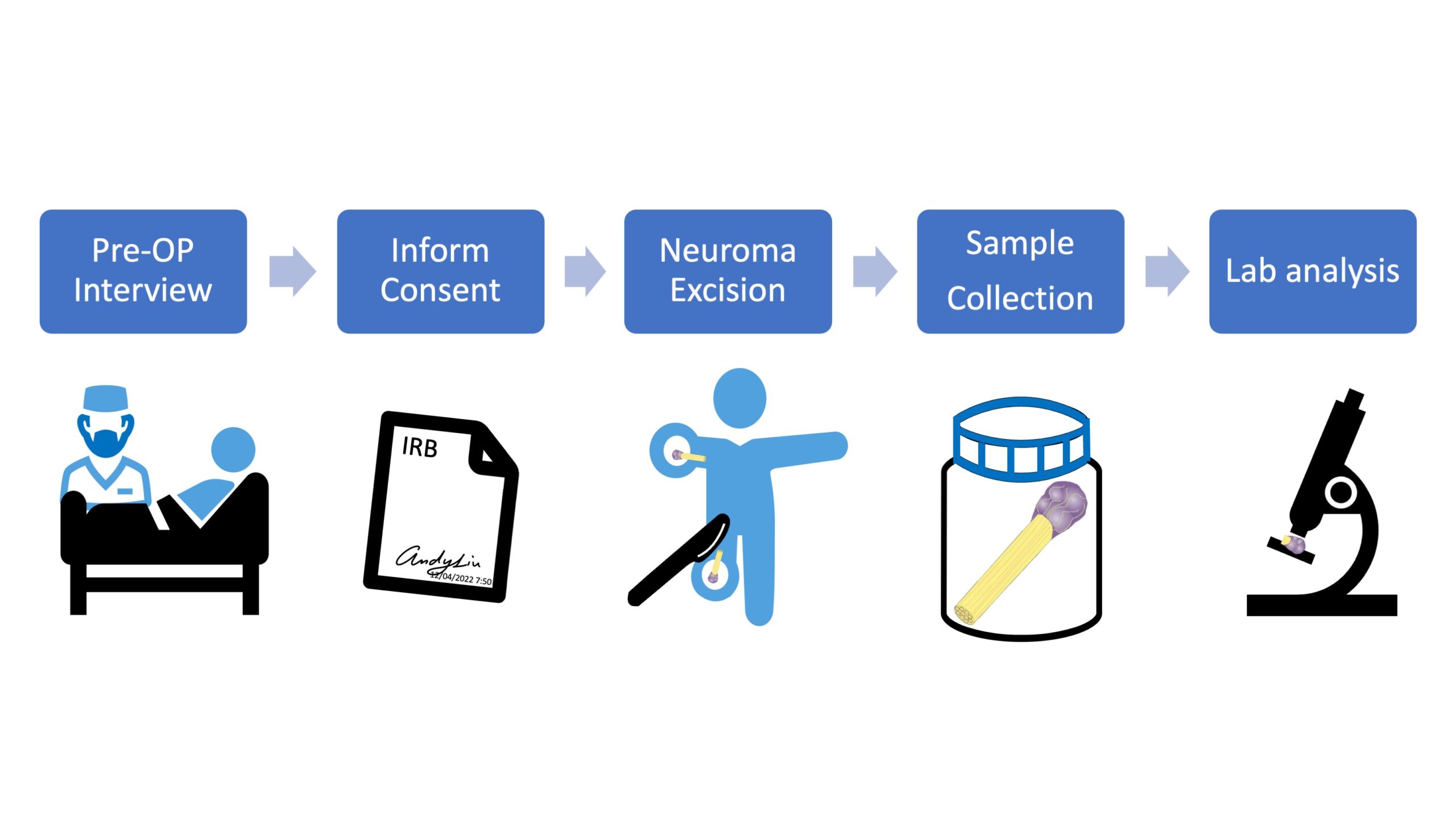
The Harvard PRECISION Human Pain Center proposes to use human TG/DRG tissue and painful neuromas to generate large-scale multi- omics datasets for the discovery and characterization of functional genetic elements, epigenetic signatures, and molecular/cellular pathways that underlie human pain transduction, transmission, and processing. As one of the key components of the Center, the Data Core aims to act as a data center by building data infrastructure, providing data management, coordination, analysis, and sharing to the investigators within the Center and other centers in the community. The projects will generate nearly 4,000 datasets of clinical (pain scores, demographics), single-cell RNA-seq, single-cell ATAC-seq, SNP genotyping, spatial transcriptomics from MERFISH, and physiology for over 1,700 samples from 150 donors.

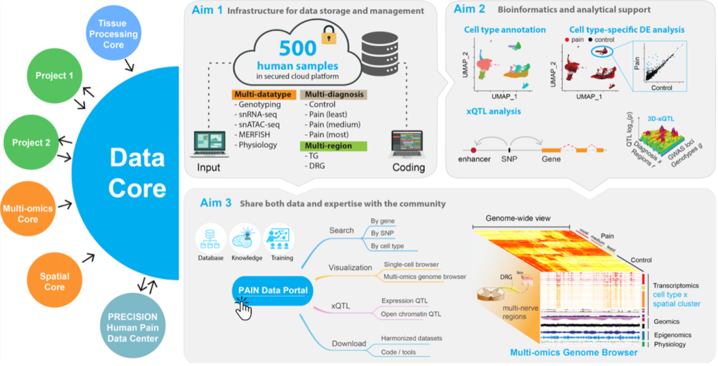
The Data Core will first harmonize the datasets and store in centralized MySQL database with accessible web interface for other cores in the Center. Standard bioinformatics pipelines will be developed to process the raw data in a systematic manner, including cell type annotation and spatial location in each brain region. The Data Core will also support the research Projects on their customized analysis, including power analysis, differential expression and chromatin accessibility across different cell types, brain regions, and diagnosis. It will also reveal the genetic variants that are associated with various molecular features via xQTL analysis. Last, all these computed blocks and tools will be integrated into a comprehensive, cloud-based, interactive web portal – Pain Data Portal, providing a generally useful resource for generating biological hypotheses, discovering effective diagnosis biomarkers, and developing new treatments for pain. The Data Core will also ensure a timely data transfer and expertise sharing with the other U19 and U24 centers in the community.

Multi-omics Core:

One of the stated objectives of the Human PRECISION Pain Network is to support multidisciplinary groups in the use of high throughput single-cell analyses on primary human tissues involved in pain processing. Toward this end, the proposed Harvard PRECISION Human Pain Center project leverages single-cell protocols that have been developed by Dr. Renthal’s laboratory to characterize human sensory ganglia. The Multi-omics Core will be led by Dr. Renthal and centralizes the single-cell transcriptomic, epigenomic, and next-generation sequencing efforts with an experienced team in both the development, optimization, and analysis of single-cell and next- generation sequencing efforts. The centralization of these cutting-edge technologies will ensure that the established protocols can be applied systematically to all samples from the Center. Moreover, it offers significant cost effectiveness as infrastructure components, including a 10X Chromium Controller, an Agilent TapeStation, and an Illumina Nextseq 550 sequencer, are already present in the proposed Multi-omics Core.

Spatial Core:

Cartoon depiction of the barcoding process used by MERFISH. Single RNA molecules are imaged and identified, in situ, by performing a series of single-molecule fluorescence in situ hybridization (FISH) experiments, with each RNA molecule identified by a fluorescence on/off pattern unique that that RNA.
The behavior of tissues arises from the interplay of the cell types and states that comprise them; the identity and function of these cells are determined by the specific fraction of the genome they express; and cellular behavior and the behavior of the tissue depend on the spatial organization of these molecules within cells and of cells within the tissue. Dissociated single-cell technologies such as single-cell RNA sequencing (scRNAseq) have revolutionized our understanding of the cellular diversity of a wide range of tissues by providing a transcriptome- wide definition of cell types and states. Unfortunately, by extracting molecules and dissociating cells before profiling expression, these methods naturally discard this essential spatial context. It is for this reason that there is a demand for technologies that can capture the benefits of transcriptome-scale profiling while preserving the essential tissue- and cellular-scale context of RNAs within intact samples.

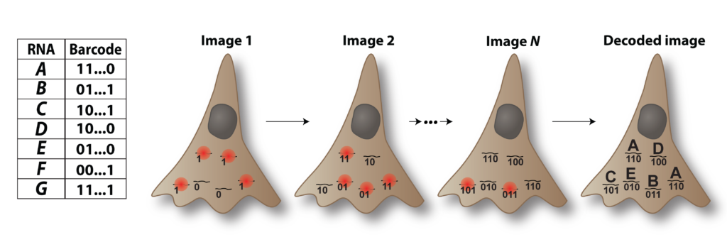
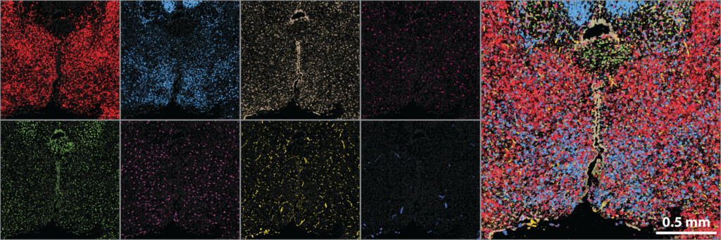
Image-based approaches to single-cell transcriptomics are the emerging and exciting answer to this demand, as these methods quantify single-cell expression profiles within fixed, intact samples via massively multiplexed, single-molecule RNA imaging. One such approach is MERFISH (multiplexed, error-robust, fluorescence in situ hybridization) which distinguishes individual RNAs with binary barcodes read out through sequential rounds of FISH staining and imaging. MERFISH has the proven ability to profile hundreds to ten thousand RNAs with near 100%- capture efficiency, single-molecule sensitivity, high throughput, and sub-micron resolution, and to leverage these capabilities to discover, define, and map cell types within the brain.

MERFISH was co-developed by Jeffrey Moffitt and is being extensively used and extended by the Moffitt laboratory. Thus, we have a unique opportunity to develop a spatial core in one of the few laboratories capable of these measurements and with the depth of expertise to help chart its use for the scientific goals of this center. Importantly, by integrating with a key laboratory in the spatial transcriptomics space, our core has access to the cutting edge of image-based transcriptomics, via MERFISH, and, critically, will continuously evaluate the emerging landscape of spatial profiling technologies, incorporating new technical capabilities as they emerge.

Atlasing the brain with MERFISH. Left: Individual tiles represent the spatial distribution of 8 different mRNAs measured in the same slice of the mouse hypothalamus. Right: All 8 RNAs plotted together. In total, this measurement profiled 150 RNAs, and MERFISH routinely measures hundreds to thousands of RNAs, providing genome-scale molecular atlases of a range of tissues.